 |
Our Facility & Services
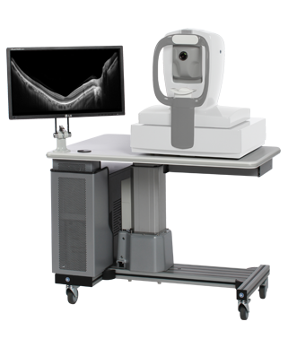
Optovue Avanti Widefield Enface OCT
|
Optical Coherence Tomography (OCT) is a standard diagnostic and imaging technique in most clinical practices today. It is a fast and noninvasive scan of the macula. By imaging the retinal histological structure, an OCT scan obtains information similar to that from an optical biopsy, but without the need for excision and histopathologic specimen processing. The OCT employs the Michelson interferometry using near-infrared light (820 nm) produced by a super luminescent diode. The light is split, and the machine compares the echo time delay of the light reflected from the retina with the echo time delay of the same light reflected from a reference mirror at a known distance. The reflected light is recombined, and the resulting interference fringe is detected and measured by a photodiode detector. The information obtained is then used to produce an image of the retina. |
Patients can expect a brief, comfortable experience with the OCT. Each scan acquisition usually takes slightly more than one second, and the entire test lasts only five to seven minutes.
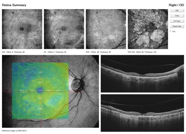
- 40° Enface OCT reference scan
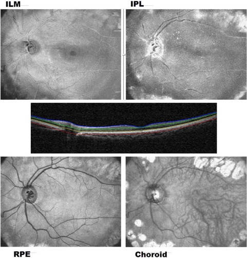
- Multi-layer Simultaneous enface assessment
- 3mm scanning depth
- Widefield scanning of high myopic eyes
- Image choroid and vitreous in a single B-scan
- High Density Cube – 320 x 320 3D Cube with SmartTM Motion Cancellation
- High-speed – Higher quality data in less time
- Real-time VTRAC Eye Tracking – higher repeatability
- High Resolution – 3µm digital
- SharpVue – unparalleled detailed images
- DCI– Deep choroid Imaging
- FLR– Fovea Location Recognition
- Advanced OCT imaging platform for future generations of clinical applications
The images can also be used to help patients understand their condition. The images give patients a clearer idea of what is going on in their eyes, and can better appreciate the effects of therapy.
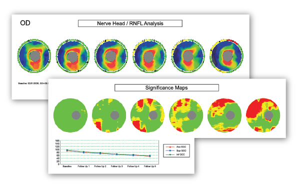
The findings of Fourier domain OCTS are objective and quantitative, and are reproducible, making documentation much more reliable.
